Studying the biomarkers of a lung cancer can change the treatment a patient receives. This is the central idea of this article and the reason why we now talk about personalized treatment.
When we diagnose lung cancer, one of the first questions we need to answer is: how far has the disease spread? But today, that question alone is no longer enough.
Two patients may have tumors that look identical on a CT scan and even under the microscope. However, their tumor cells may have different molecular alterations. This difference is not merely academic: it can mean that the most appropriate treatment for one patient is different from the treatment recommended for the other.
That is why there is no longer just one type of lung cancer, but many different types of lung cancer. And the key question has shifted from “What treatment is used for lung cancer?” to:
What is the most appropriate treatment for this specific lung cancer and this particular patient?
WHAT IS A BIOMARKER?
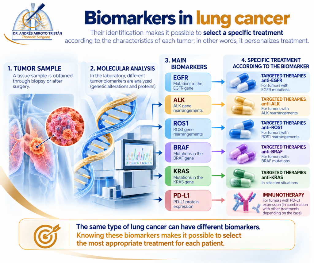
A biomarker is a biological characteristic of the tumor itself that can be analyzed in a laboratory. It may be an alteration in a gene or the presence of a protein in tumor cells. Biomarkers are primarily studied using a tissue sample and, in some cases, a blood sample (known as a liquid biopsy).
To understand their value, it is useful to distinguish between two different questions:
Staging (TNM system) answers Where is the cancer, and how far has it spread? It assesses the tumor (T), lymph nodes (N), and metastases (M).
Biomarkers answer What are the biological characteristics of this tumor?
Both types of information are complementary. Staging helps determine whether surgery, radiotherapy, systemic treatment, or a combination of these approaches is appropriate. Biomarkers help identify which specific systemic treatment may be most effective for that particular tumor.
You have probably heard terms such as EGFR, ALK, ROS1, BRAF, KRAS, or PD-L1. As a patient, you do not need to memorize them. What matters is understanding why identifying these biomarkers is important.
ONE BIOMARKER, ONE SPECIFIC TREATMENT
This is the most important concept: certain biomarkers make it possible to select a treatment specifically designed to target the alteration present in that tumor. This is the foundation of precision medicine.
When molecular analysis identifies a specific alteration, it may become a therapeutic target: a vulnerability in the tumor that can be targeted by a particular drug. These treatments are known as targeted therapies. Other markers, such as PD-L1, help assess the potential benefit of immunotherapy, which stimulates the immune system to recognize and attack cancer cells.
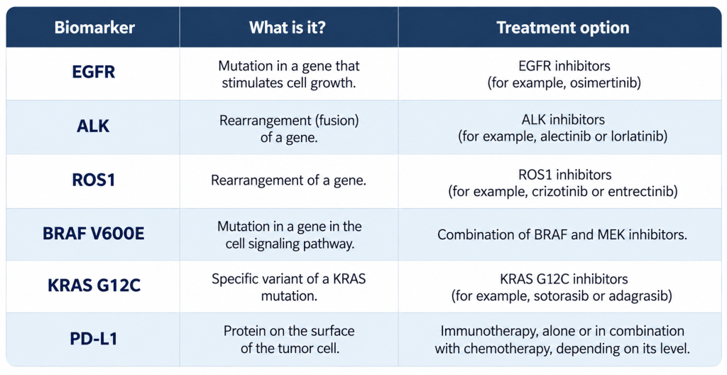
There are other molecular alterations that can be targeted with specific treatments (including MET, RET, NTRK, and HER2, among others), and the list continues to grow every year. The final choice of medication is made by the Oncology team based on the stage of the disease, the patient’s condition, and approved treatment indications.
Biomarkers do not only indicate which treatments may work. They also help identify which treatments may be less effective. For example, tumors with EGFR or ALK alterations generally respond less well to immunotherapy, making targeted therapy the preferred option in these cases. Identifying the biomarker helps avoid less effective treatments and opens the door to the most appropriate therapy. This is precisely what personalized treatment means.
SURGERY AND BIOMARKERS: A UNIFIED STRATEGY
Surgery and the molecular analysis of cancer are often considered two separate fields. In reality, they are part of the same strategy, and the thoracic surgeon plays a key role in both.
Studying biomarkers requires a sufficient quantity of high-quality tumor tissue. This tissue may come from a diagnostic biopsy or from the surgical specimen obtained when the tumor is removed.
When we perform a lung resection for cancer, the pathologist can examine the tumor in detail, including its exact type, size, and microscopic characteristics. In addition, during the procedure, we perform a lymphadenectomy, which involves removing lymph nodes from different stations within the chest for microscopic examination.
This provides us with two types of information from a single surgical procedure:
Pathological TNM staging, which is more accurate than the clinical TNM staging estimated before surgery using CT and PET-CT scans. A lymph node that appeared normal on imaging may contain microscopic cancer cells.
The tumor’s molecular profile, when molecular testing is indicated, obtained from the surgically removed tissue.
In other words, surgery helps answer two questions at the same time: how far the cancer has spread and what its biological characteristics are.
IF MY TUMOR HAS BEEN REMOVED, WHY DO I NEED FURTHER TREATMENT?
This is one of the questions we hear most frequently during consultations after surgery. The answer is that completely removing the visible tumor does not always mean that treatment is finished, and this is where biomarkers also play a decisive role.
After surgery, we know the definitive pathological stage and can better estimate the risk of microscopic cancer cells remaining outside the area that was removed. At this point, the tumor’s molecular profile can help determine which additional treatment is most appropriate:
In patients who have undergone surgery and whose tumors have an EGFR mutation, targeted treatment after surgery has been shown to reduce the risk of cancer recurrence in certain stages.
For surgically removed tumors with an ALK rearrangement, additional targeted treatment after surgery is also available for selected stages.
Before considering immunotherapy either before or after surgery, it is important to determine whether the tumor has EGFR or ALK alterations, as these can change the treatment strategy.
This is why biomarker testing is no longer reserved exclusively for advanced disease. Even in operable lung cancer, identifying biomarkers can change the treatment approach. The available options (chemotherapy, radiotherapy, immunotherapy, targeted therapies, or combinations of these treatments) are determined individually by a multidisciplinary tumor board.
THE KEY TAKEAWAY
We no longer treat just an organ called the lung or an image on a CT scan. We treat an individual patient with a specific tumor. To do this, we consider the type of tumor, how far it has spread (TNM staging), and, when indicated, its biomarkers. Thoracic surgeons, oncologists, radiation oncologists, pulmonologists, radiologists, nuclear medicine specialists, and pathologists work together to analyze this information.
If you or a family member has been diagnosed with lung cancer, the following questions may be helpful during your consultation:
Has my tumor’s biomarker profile been analyzed?
Is there enough tissue to perform this analysis, or do I need another biopsy?
Does my tumor have any molecular alterations that can be targeted with specific treatments?
How do these results affect my treatment, before or after surgery?
Knowing where the tumor is located remains essential. But understanding its biological characteristics is what allows us to choose a specific, personalized treatment for each patient.
This article is for informational purposes only and does not replace an individual medical assessment by your healthcare team.